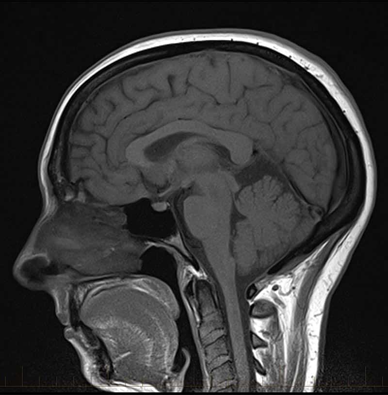

The Image Feature Detection Flask application offers a web-based interface for performing advanced feature detection on a pre-set collection of images. It integrates the robust computer vision capabilities of OpenCV to apply different feature detection algorithms, providing users with immediate visual feedback on the results of various image analysis techniques.
The application simplifies the process of exploring computer vision algorithms, offering a user-friendly interface suitable for educational purposes and quick demonstrations of feature detection technology.
The app is deployed on a Flask server and accessible via a web browser, ready to demonstrate feature detection with minimal setup.
To use the application, users can select an image from the dropdown menu and choose a feature detection algorithm. Upon selection, the application processes the image using the chosen algorithm and displays the results in real-time. This immediate feedback allows users to observe the impact of different algorithms on the same image, facilitating comparisons and learning. The processed image is stored for future reference, enabling users to revisit previous results without reprocessing the image. This efficient workflow enhances the user experience and encourages exploration of various feature detection techniques.
Future updates may include user image uploads, additional algorithms, and further optimizations to enhance the application’s interactivity and educational value.
Region of Interest (ROI) Selection: To provide users with more precise analysis capabilities, we plan to implement a Region of Interest selection tool within the application. This feature will allow users to specify particular areas of the image for focused feature detection. By simply clicking and dragging over the desired area, users can define the ROI, which can then be used to apply feature detection algorithms exclusively within this region. This is particularly useful in scenarios where users are interested in analyzing specific parts of an image, such as identifying features in a particular object or segmenting a specific structure within a medical image. Implementing ROI selection can enhance the application's utility significantly, especially for users who deal with complex images where global feature detection might yield too much noise. In medical imaging, for instance, focusing on a tumor or a specific brain region can provide more meaningful insights than processing the entire image. ROI selection could be accompanied by additional tools for adjusting the size and shape of the selected area, as well as the ability to move the ROI around the image for comparative analysis. With this enhanced control, the application could serve an even broader range of use cases and provide more value to researchers, clinicians, and hobbyists interested in targeted image analysis. In terms of technical implementation, ROI selection can be achieved using client-side scripting, where mouse events are used to draw a rectangle over the image. This rectangle's coordinates can then be sent to the server-side Flask application, where OpenCV can process only the selected image region. Adding this feature would certainly make the application more powerful and versatile for users requiring detailed image analysis capabilities.
Optimization for Varied Image Types: Recognizing the diverse needs of our user base, we aim to optimize the application for a variety of image types beyond standard photographs. One such advancement will target the analysis of EEG signal images, focusing on the detection of seizure spikes, a critical aspect of neurological diagnostics. Seizure Spike Detection in EEG Signals: EEG (electroencephalogram) signals provide a wealth of information about brain activity and are crucial in diagnosing and monitoring neurological disorders like epilepsy. Our planned feature will introduce algorithms specifically tuned to identify seizure-related anomalies in EEG images. By applying specialized filtering and feature detection techniques, we can assist healthcare professionals in rapidly pinpointing areas of interest in EEG data. Custom Algorithm Development: This will involve developing or integrating custom algorithms tailored for the unique patterns and characteristics of EEG signals. We will explore machine learning approaches, particularly in the realm of convolutional neural networks (CNNs), which are adept at recognizing patterns in visual data. User-Defined Parameter Tuning: To accommodate the highly specialized nature of EEG analysis, we will provide interfaces for users to adjust algorithm parameters. This flexibility will enable professionals to calibrate the feature detection to their specific requirements, potentially improving the accuracy of seizure spike detection. Collaboration with Medical Professionals: In developing these features, we will seek partnerships with medical practitioners and researchers. Their expertise is invaluable in fine-tuning the application to meet clinical standards and ensure that the tool provides practical benefits in a medical setting. Educational and Research Use: Beyond clinical applications, this feature could serve as an educational tool for students and researchers interested in biomedical signal processing. It would provide an accessible way to visualize and understand the complexities of EEG signals and seizure activities. Incorporating such specialized feature detection would not only broaden the application's scope but also deepen its impact, especially in critical fields such as medical imaging and research. The ability to process and analyze EEG signals effectively could turn the application into a powerful tool for both educational and diagnostic purposes.